Analyse et visualisation de volumes des glandes sébacées

La microtomographie à rayons-X, véritable microscopie 3D non destructive, permet de s’affranchir des limitations des microscopies 2D.  Novitom utilise la microtomographie de manière experte pour étudier la structure interne des explants de peau, par exemple pour caractériser les glandes sébacées. Il est ainsi possible de les visualiser tout en laissant l’explant dans son milieu de conservation, de mesurer leur surface et volume dans l’ensemble de l’explant par analyse d’images 3D, ou réaliser des coupes virtuelles à l’intérieur de ce volume.

What are the assets of synchrotron X-ray microtomography ?

++ Non-invasive, real-time analysis

SR-μCT allows direct, non-invasive monitoring of structural changes in pellets, in particular the formation of cracks in polymeric coatings, without complex sample preparation. This reduces the risk of creating artefacts and provides a more accurate analysis of the internal dynamics of solid pharmaceutical dosage forms.

++ Optimisation of formulations

By better understanding the underlying mechanisms of drug release, such as osmosis and mechanical stress, manufacturers can optimise the design of coated tablets, reducing the amount of trial and error usually required in the development of new formulations.

++ Improved modelling and prediction

The data obtained from SR-μCT is also used to develop models, making it easier to predict drug release behaviour as a function of the composition of coatings and the internal structure of tablets, lozenges or pills.

As an independent lab, Novitom enables companies to access synchrotron micro and nano-tomography :

  • Regular access to instruments at synchrotron facilities
  • Know-how in experimental design for real-time microtomography
  • Expertise in extracting information and physical parameters from raw data and in combination with ancillary techniques

Autres articles de blog xx

Revendications pour le renforcement des propriétés mécaniques

Revendications pour le renforcement des propriétés mécaniques

La technique de microscopie à forme atomique est utilisée pour réaliser une topologie (profilomètrie) de la cuticule tout en mesurant…

Revendication pour la pénétration d’un actif dans la cuticule

Revendication pour la pénétration d’un actif dans la cuticule

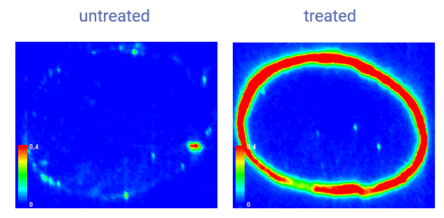
Observer et quantifier la présence d’un actif dans un tissu est souvent difficile à cause des très faibles quantités de…
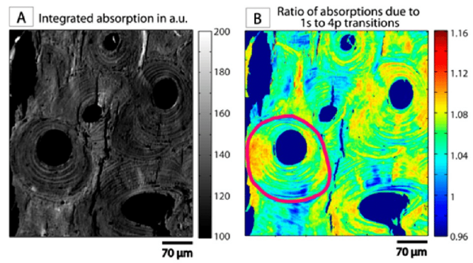
Analyse et visualisation de volumes des glandes sébacées

Analyse et visualisation de volumes des glandes sébacées

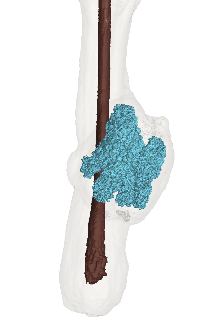
La microtomographie à rayons-X, véritable microscopie 3D non destructive,…

The dissolution of coated tablets highlighted by X-ray microtomography

The dissolution of coated tablets highlighted by X-ray microtomography

Initial question Which phenomena are decisive in oral drug…