Spectroscopie d’absorption des rayons X du calcium à l’échelle du micron sur l’os cortical
Spectroscopie de l’absorption des rayons X de l’angle K du calcium à l’échelle du micron sur l’os cortical : Les…
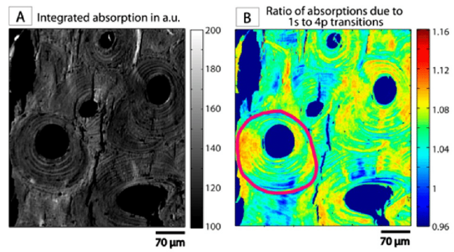
Spectroscopie de l’absorption des rayons X de l’angle K du calcium à l’échelle du micron sur l’os cortical : Les…
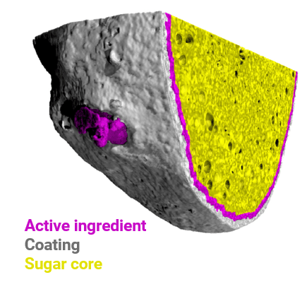
La microtomographie à rayons-X, véritable microscopie 3D non destructive, permet de s’affranchir des limitations des microscopies 2D. Novitom utilise la…
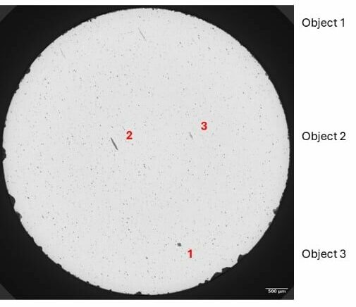
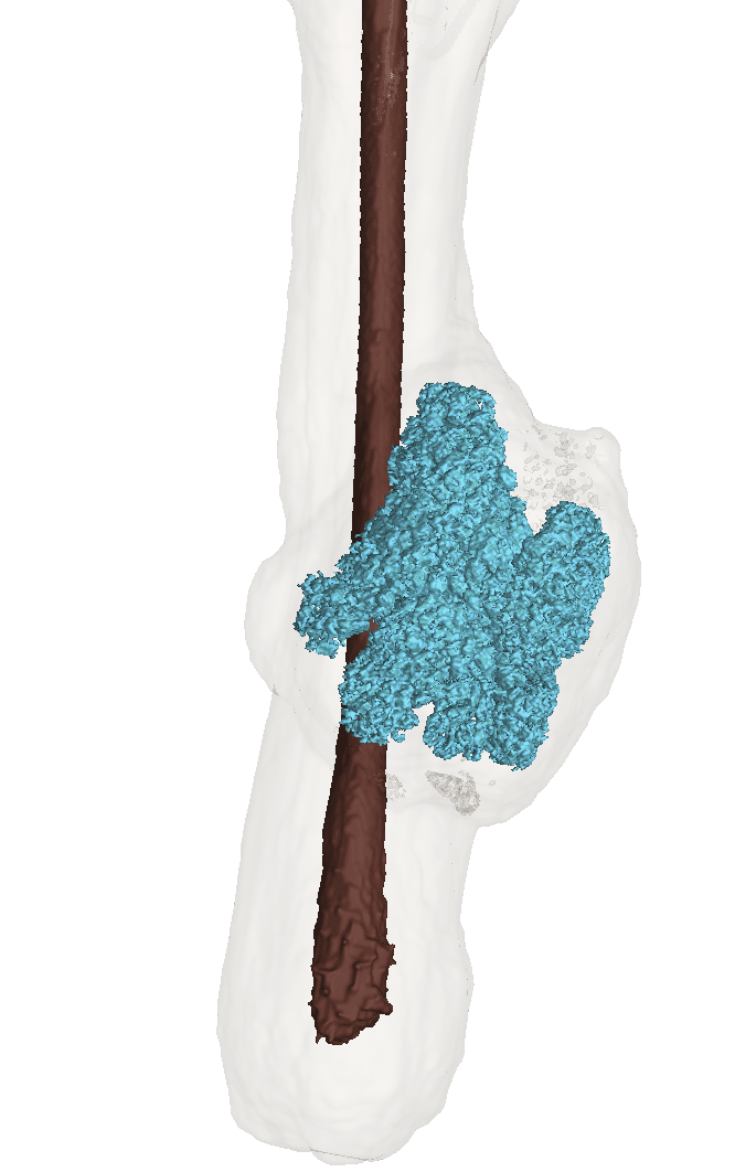
Afin d’étudier la rupture de pièces critiques comme peuvent l’être les aubes de turbine, la morphologie des inclusions de différents…
Spectroscopie de l’absorption des rayons X de l’angle K…
La microtomographie à rayons-X, véritable microscopie 3D non destructive,…